Mechanical asphyxia medico-legal. In forensic medicine, mechanical asphyxia is understood as acute asphyxia, which occurs as a result of partial or complete cessation of air access into and into the lungs, caused by various mechanical obstacles. Depending on the nature of the mechanical factors that create obstacles to breathing, and the characteristics of their impact, A. m. is distinguished from compression of the neck - strangulation asphyxia (strangulation with a noose, strangulation with hands); A. m. from compression of the chest and abdomen - compression asphyxia; A. m. from closing the openings of the nose and mouth with soft objects, closing the airways with foreign bodies, liquids - obstructive asphyxia; A. m. from the closure of the respiratory tract with food masses and blood. As a rule, A. m. proceeds acutely, ending in death (if not interrupted) within 6-8 min. In severely weakened individuals, in patients with, for example, coronary heart disease, it may occur in the first minutes. The pathomorphological changes found in A. m. are based on acute circulatory disorders. Their manifestations, which are called general signs of asphyxia, are not pathognomonic for A. m., since they can also occur in other types of quickly occurring death with a similar genesis. The general signs of asphyxia revealed during autopsy include abundant, diffuse, intensely colored, appearing after 40-60 min after death; skin of the face and neck; pinpoint hemorrhages in the conjunctiva; involuntary urination, pushing out the mucus plug from the cervical canal; dark liquid in the heart and large venous vessels, overflow of blood from the right half of the heart compared to the left, venous congestion of the internal organs; subpleural and subepicardial hemorrhages (Tardier spots). In addition to the general signs, with each type of A. m., specific manifestations arise, which serve as the basis for expert evidence of death from asphyxia and the specification of its type. for example, the most typical external signs of hanging are strangulation (a mark on the skin from compression by a noose, rice. 1
), width, depth, bottom relief, density and which depend on the material of the loop and the degree of post-mortem drying of the skin; severe bluishness of the facial skin; the tip of the tongue protruding from the mouth, usually clamped between the teeth (bitten); subconjunctival hemorrhages; traces of urination and defecation, ejaculation. In a typical oblique-ascending position, the loop is located anteriorly in the upper part of the neck or at the level of the thyroid cartilage. There is also a post-mortem application of a noose in order to conceal a crime by simulating hanging. Therefore, in each case, the expert must resolve the issue of the lifetime of the formation of the strangulation furrow. This is evidenced by hemorrhages in the skin along the periphery of the groove, in the marginal and intermediate ridges (if the groove is not single), in the subcutaneous tissue and it, as well as in the soft tissue around fractures of the cartilage of the larynx and sublingual, which are sometimes observed during hanging. In regional lymph nodes and small vessels of the lungs, small fat emboli from crushed subcutaneous tissue along the strangulation groove can sometimes be found. In the opened carotid arteries, transverse tears in the intima may be detected (Amusse's sign, rice. 2
), and in the lungs - pinpoint subpleural hemorrhages (Tardier spots, rice. 3
). Asphyxia from compression of the chest and abdomen occurs as a result of a sharp difficulty and cessation of respiratory movements of the chest (during collapses in mines, sand and gravel quarries, in a crowd, etc.). In addition to general signs, when examining the corpses of people who died from this type of A. m., puffiness and severe cyanosis of the face with multiple small hemorrhages in the skin (), as well as the so-called carmine pulmonary edema, are found. In some cases, if the abdomen is compressed by blunt hard objects of large mass, bones, soft tissues and internal organs may appear. Evidence of death from asphyxia as a result of obstruction of the respiratory tract by foreign bodies, food masses, and granular substances is their detection in the respiratory tract. The level of obstruction depends on the caliber of foreign matter, for example, food masses and small granular substances penetrate the respiratory tract up to the bronchioles and alveoli. Asphyxia can be caused by the airway being blocked by soft objects or another person's hands. Moreover, if the victim could not offer resistance, then no specific signs of this type of asphyxia are detected. Sometimes, when soft objects are strongly pressed to the nose and mouth, a trace of the texture of the fabric may remain on the skin: abrasions and bruises are possible on the mucous membrane of the lips and gums. When examining the corpses of persons who died from asphyxia, the tasks of the forensic expert include establishing the type of asphyxia, its lifetime, thanatogenesis and causes, and the duration of death. When examining persons who have undergone A. m., it is necessary to establish its very fact, asphyxia and the resulting consequences. Bibliography: Avdeev A.I. Forensic medical corpse, M., 1976; Botezatu G.A. and Muthoi G.L. Asphyxia, Chisinau, 1983, bibliogr.; , ed. A.A. Matysheva and A.R. Denkovsky, L., 1985; Judicial, ed. V.M. Smolyaninov. With. 67, M., 1982.

1. Small medical encyclopedia. - M.: Medical encyclopedia. 1991-96 2. First aid. - M.: Great Russian Encyclopedia. 1994 3. Encyclopedic Dictionary of Medical Terms. - M.: Soviet Encyclopedia. - 1982-1984.
See what “mechanical asphyxia” is in other dictionaries:
A., caused by a mechanical obstruction to breathing (obstruction of the respiratory openings and tracts, compression of the neck, chest and abdomen) ... Large medical dictionary
ASPHYXIA- – a state of increasing suffocation, leading to a lack of oxygen in the blood and tissues (hypoxia) and the accumulation of carbon dioxide in them (hypercapnia). The main causes of asphyxia: 1) compression of the upper respiratory tract from the outside during hanging, strangulation... ... Encyclopedic Dictionary of Psychology and Pedagogy
ASPHYXIA- – suffocation caused by the cessation or sharp reduction of oxygen supply to the body. In some cases, obstacles to oxygen access can be external and purely mechanically impede breathing, in others, interstitial metabolism is disrupted... ... Soviet legal dictionary
I Poisoning (acute) Poisoning is a disease that develops as a result of exogenous exposure to the human or animal body of chemical compounds in quantities that cause disturbances in physiological functions and pose a danger to life. IN … Medical encyclopedia
PREMATURE- (praematuritas, Friih geburt), in the usual sense, the birth of a child before the full term of pregnancy. This definition, however, is theoretically and practically unsatisfactory, because firstly the moment of conception, i.e. the beginning and therefore... ... Great Medical Encyclopedia
Large medical dictionary
- (Latin strangulatio strangulation; Greek strangule noose, gallows) mechanical asphyxia caused by compression of the neck, for example. loop, hands... Large medical dictionary
A category of forensic medical classification of death, identified on the basis of the nature of the direct external influence that caused violent death (mechanical asphyxia, trauma, poisoning, etc.), or the immediate cause... ... Medical encyclopedia
- (ZOMP) a complex of organizational, engineering, medical and other measures aimed at preventing or maximally weakening the damaging and destructive effects of nuclear, chemical and biological weapons with the aim of... ... Medical encyclopedia
- (Latin strangulatio strangulation; Greek strangulē noose, gallows) mechanical asphyxia caused by compression of the neck, for example with a noose, by hands... Medical encyclopedia
I Tetania (tetania; Greek tetanos tension, spasm) is a pathological condition characterized by convulsive syndrome and increased neuromuscular excitability due to a decrease in the concentration of ionized calcium in the blood serum, as... ... Medical encyclopedia
Asphyxia is a pathological condition that develops acutely and disrupts the functions of vital systems of the body. It occurs due to a sharp decrease in oxygen supply to the organs. Insufficient gas exchange between the external environment and the body leads to the accumulation of carbon dioxide in the tissues. Oxygen starvation and the inability to breathe normally ends in loss of consciousness and death. Death as a result of asphyxia can also occur due to reflex arrest of the heart muscle. A similar condition is diagnosed when the superior laryngeal nerve is irritated from compression of the neck.
Signs of asphyxia
Numerous post-mortem signs are determined by the rate of death, the characteristics of the body and the lifetime course of suffocation. They are also present in other variants of quick death. Among them there is not a single one that is constant and absolutely true. External and internal signs of death from asphyxia are determined.
Internal signs
Choking is diagnosed by a number of clinical symptoms. The color and coagulability of the blood matters. After death, the blood darkens due to its conversion from arterial to venous due to the rapid absorption of red blood cells into tissues.
Liquid blood is a common sign of rapid death. Explained by saturation with carbon dioxide, autolysis. Blood clots are observed rarely, with a slow course of asphyxia. Coagulation is associated with leukocytosis, but with rapid death it is not present.

Point hemorrhages, or Tardieu spots under the membrane of organs, are considered an indicator of death. They arise due to increased permeability of the walls of blood vessels and rupture of capillaries. Other internal signs include congestion of organs, respiratory tract mucosa, blood overflow of the right atrium and ventricle, anemia of the spleen. These symptoms can be observed not only with suffocation.
External signs
External signs of death from lack of oxygen include: They have an intense blue-purple color. Appear due to the movement of a large amount of blood to the lower parts of the body. The color is caused by oxygen-poor and carbon dioxide-rich blood.

Death from asphyxia is indicated by cyanosis of the face and nails. It is observed in the first stage of suffocation. The reason is blood stagnation, expansion and overflow of the blood vessels of the head. The cyanosis disappears within a few hours after death. The pathological process is accompanied by involuntary urination and.
Causes of asphyxia
The reasons come down to two groups. The first is characterized by a disorder of external respiration, the second - interstitial. Suffocation can occur in the absolute absence of oxygen while in a closed space. Common causes of choking include:
- mechanical compression of the neck, chest, abdomen;
- respiratory tract damage;
- their closure with liquid or a foreign body;
- accumulation of air or blood in the pleural cavity due to injury;
- cooling;
- poisoning.
Asphyxia is the cause of death due to electric shock. It is also observed in infectious processes, epilepsy, accompanied by spasm of the respiratory muscles. Suffocation is caused by dysfunction of the respiratory center, which occurs due to organic damage. This outcome is observed when

Attention! The oxygen reserve in the body is 2-2.5 liters. The mentioned volume is only enough to preserve life for several minutes.
Suffocation occurs during high-altitude hypoxia. Poisoning with strychnine and other substances can also be aggravated by asphyxia, convulsions, and death.
Asphyxia Clinic
The main sign of suffocation is failure of breathing. Develops gradually, paroxysmally or suddenly. In acute asphyxia, breathing becomes frequent, deep and noisy. Inhalations are longer than exhalations. The reason is irritation of the respiratory center by carbon dioxide. The auxiliary muscles are included in the respiratory act, and the intercostal spaces and epigastric region recede.
The skin takes on a blue-purple coloration on the face and neck. The period of excitement is replaced by increasing muscle weakness and slowing of the heartbeat. In the first minute, loss of consciousness occurs. After breathing and cardiac activity stop, death occurs.
Types of asphyxia
Strangulation can be intrauterine, primary or secondary. The first two types include asphyxia of the fetus and newborn. Secondary asphyxia includes:
- mechanical asphyxia;
- reflex asphyxia;
- suffocation from lack of oxygen in the air;
- asphyxia due to damage to the nervous system;
- asphyxia, which develops with spasticity.

Death from mechanical asphyxia is more often diagnosed. This type of strangulation occurs due to compression of the neck with hard objects and through hanging, strangulation with hands or a noose. Occurs when the chest and abdomen are compressed (compression asphyxia). Varieties include drowning, blocking of the respiratory tract with foreign bodies, and suffocation with vomit. Hanging and drowning account for the largest percentage.
When examining the corpse, general signs of death from mechanical asphyxia are revealed. These include cyanosis of the facial skin, slow cooling of the body, involuntary defecation, urination, ejaculation, moderate. The sign is pinpoint hemorrhages in the conjunctiva of the eyelids.
Stages of suffocation
Regardless of the specifics of the factors that initiate strangulation, a distinction is made between pre-asphyxial and asphyxial periods of its development. The first period lasts from 10 seconds to 1 minute, the second is conditionally divided into successive stages.
|
Stage |
Clinical course |
| Stage of inspiratory dyspnea |
|
| Stage of expiratory dyspnea |
|
| Stage of short-term respiratory arrest |
|
| Terminal respiration stage |
|
| Persistent respiratory arrest |
|
The duration of the pathological process is 5-6 minutes. After this time, irreversible changes occur in the cerebral cortex. The duration of the stages is influenced by the person’s age, health, and type of suffocation.
The task in case of suffocation is to quickly restore normal functioning of the respiratory tract. The preservation of the life and health of the victim depends on the speed of action. You should call and see a doctor.

Algorithm for providing emergency care:
- If a person is conscious, but cannot breathe due to a foreign body in the airway, you need to stand behind him and put your arms around his waist.
- Make a fist with one hand. Clasp your fist with your other hand.
- With a sharp movement, press on the stomach below the ribs above the navel.
- Repeat the steps until the object comes out of the respiratory tract.
Providing assistance in each case has its own specifics and depends on the reasons that led to strangulation. Thus, the supply of oxygen contained in the human body is negligible. Acute oxygen starvation of tissues leads to disruption of metabolic processes at the cellular level and death of the body.
Video
Mechanical asphyxia - disturbance of external respiration as a result of mechanical impact.
Damaging factor - a mechanical effect that prevents air from entering the lungs and causes hypoxia and hypercapnia.
During mechanical asphyxia, several periods are distinguished.
1. Pre-Asphyctic period begins when the flow of air into the lungs ceases.
2. The period of inspiratory dyspnea. Lack of oxygen in the body activates the inspiratory section of the respiratory center of the medulla oblongata and leads to inspiratory dyspnea. There is a loss of consciousness and severe physical inactivity, which exclude self-rescue.
3. The period of expiratory dyspnea. The accumulation of carbon dioxide in the body leads to activation of the expiratory part of the respiratory center of the medulla oblongata and expiratory shortness of breath. Excitation of smooth muscles combined with relaxation of the sphincters leads to involuntary defecation, urination and ejaculation.
4. A period of short-term cessation of breathing. The accumulation of carbon dioxide leads to a complete cessation of the functioning of the respiratory center of the medulla oblongata and cessation of breathing.
5. The period of terminal respiration. Continued hypercapnia stimulates the respiratory center of the spinal cord. The breathing movements are short and slurping.
6. The period of final cessation of breathing. Excess carbon dioxide leads to exhaustion and paralysis of the respiratory center of the spinal cord. Irritation of the center of the vagus nerves, a decrease in blood pressure and a slowdown, and then a drop in cardiac activity, complete the course of the asphyxial process. Single heartbeats after the final stop of breathing are observed for 5-30 minutes.
With mechanical asphyxia, an autopsy reveals general signs of acute death, as well as specific signs indicating its various types.
General signs of asphyxia. In asphyxia that ends in death, a number of general morphological signs are usually observed, called general asphyxia, or signs of quickly occurring death:
Facial cyanosis, often with ecchymosis;
Pinpoint hemorrhages in the conjunctiva of the eyelids, most densely located in the transitional folds;
Intense diffuse purple-bluish cadaveric spots with multiple intradermal hemorrhages (cadaveric ecchymoses);
Involuntary defecation, urination and ejaculation;
Dark shade of blood;
Venous congestion of internal organs;
Acute swelling of the lungs;
Overflow of blood from the right side of the heart;
Small focal hemorrhages under the visceral pleura and epicardium (Tardier spots);
Liquid state of blood.
Particular signs of asphyxia reflect some specific signs indicating the mechanism of development of the asphyxial process. Depending on the mechanism of occurrence, it is customary to distinguish four types of mechanical asphyxia: strangulation, obstruction, compression and asphyxia in a confined space.
1. Strangulation asphyxia- compression of the neck organs by a noose, a traumatic object or body parts of an unarmed person. With strangulation asphyxia, a lesion is formed on the neck, which is called a strangulation groove.
Forensic significance of the strangulation furrow Determining the type and material of the loop or traumatic object (Fig. 26). A closed loop surrounds the neck on all sides, forming a closed strangulation groove. Blunt hard and soft flexible objects (loops) form closed and open strangulation grooves. Hard loops form deep, dense furrows with clear edges and pronounced settling; soft ones leave a mark in the form of a depressed strip-like area of intact skin
gray color. Single, double or multi-turn strangulation grooves arise from the action of single, double or multi-turn loops. Multi-turn furrows are separated along the length by an intermediate roller. Blunt, hard, inflexible objects form open and atypical open strangulation grooves. An atypical open strangulation groove is formed from the fork of a tree, the back of a chair, the crossbar of a stool, or a part of the body of an unarmed person (hand, forearm, shoulder and forearm, knee joint area, lower leg, foot, etc.). Microparticles of the loop material are often fixed on the surface of the strangulation groove.
Determination of the surface relief of a traumatic object. The width of the groove corresponds to the width of the loop. The twisted rope leaves a mark in the form of oblique parallel striped impressions; waist belt - continuous sedimentation, twisted sheet or towel - intermittent thin elongated narrow intersecting strips of intradermal hemorrhages located longitudinally or obliquely relative to the length of the groove. The action of the node leads to local pinching of the skin with multiple intradermal hemorrhages, limited sedimentation, the shape and size of which may correspond to the shape and size of the node.
Determination of intravital or postmortem formation of strangulation furrow. The intravital formation of the groove is evidenced by sedimentation and intradermal hemorrhages along the strangulation groove, hemorrhages in the subcutaneous tissue and neck muscles in the projection of the strangulation groove, hemorrhages in the legs of the pectoral muscles, in the area of tears in the intima of the carotid arteries, in the pharynx, root and in the area of pinched tissue of the tongue, retrobulbar fiber, back muscles, intervertebral discs, legs
Rice. 26. Hard loops form deep grooves with clear edges, soft loops leave a mark in the form of a depressed area of intact skin
diaphragm, in regional lymph nodes, hemorrhages around fractures of the hyoid bone and cartilage of the larynx, swelling of the subcutaneous tissue of the neck above the level of the strangulation groove, bleeding from the nose, mouth and ears, reactive changes in the area of hemorrhages, changes in the tinctorial properties of the skin, impaired activity of a number of enzymes and necrobiotic changes in muscle fibers in the pressure band, detected by histological and histochemical research methods. Postmortem formation of the groove is indicated by the absence of signs of intravital strangulation groove.
Conditions for the occurrence of strangulation asphyxia
Hanging - a type of strangulation asphyxia, which occurs from compression of the neck organs by a noose tightened under the weight of the body of the deceased. Hanging is most often a suicide, although accidents, murders, and execution by hanging occur. When hanging, the strangulation groove is not closed (the intact part of the neck corresponds to the position of the free end), unevenly depressed (on the surface of the neck opposite the node, the groove is most pronounced), located in the upper third of the neck (above the thyroid cartilage) in an obliquely ascending direction (Fig. 27) .
When hanging in a sitting or lying position, the location of the walking groove changes, however, the open and uneven nature of the groove will still indicate hanging. When hanging in a sitting position, two strangulation grooves may form as a result of the displacement of a loose loop due to body sliding. When hanging in a confined space, it is possible that superficial damage may occur in the agonal period, during convulsions from impacts on nearby objects. If a corpse touches hot objects (heating radiator), not only superficial but also deep contact burns can occur on the body.
To indirect signs indicating longitudinal stretching

Rice. 27. Hanging: strangulation groove is intermittent, unevenly depressed, obliquely ascending from front to back, located in the upper third of the neck
neck pain include: transverse ruptures of the intima of the common carotid arteries (Amousse’s sign); hemorrhages in the adventitia of these vessels (Martin's sign) and in the medial legs of the sternocleidomastoid muscles; bleeding from the nose, ear and mouth; infringement of the tongue; Friberg's sign (local vacuolization of the intima of the carotid arteries); hemorrhages in regional lymph nodes; damage to the bones and cartilage of the larynx; fracture of the odontoid process of the second cervical vertebra; hemorrhages in the intervertebral discs (Simon's symptom) and ruptures of the ligaments fixing the cervical spine; damage to the cervical spinal cord; hemorrhages in the postorbital tissue, in the root and tissue of the tongue, pharynx; the formation of cadaveric spots in the lower extremities and hands (with a long stay of the corpse in a loop in a vertical position); lengthening of the neck and sometimes complete separation of the body.
Loop removal - a type of strangulation asphyxia that occurs when the neck is evenly and tightly compressed by a loop. Almost always the noose is tightened by an outside hand. When compressed with a loop, the strangulation groove is located horizontally, at the level of or below the thyroid cartilage, evenly depressed, and has a closed character. When various objects fall under the loop, the strangulation furrow will not be closed, but throughout the rest of the length it will be expressed generally evenly. One of its sections, corresponding to the position of the twist, knot or overlap of the free ends, as a result of pinching the skin, will have multiple mutually intersecting short and narrow stripes of intradermal hemorrhages or linear abrasions (Fig. 28).
Hand strangulation - a type of strangulation asphyxia that occurs when the neck is compressed by the fingers or between the forearm and shoulder. From the action of the fingers on the neck, round or oval bruises appear, located at a short distance from each other, in groups of two to four. Against the background of bruises, arched or short strip-like bruises sometimes form.

Rice. 28. Loop removal: the strangulation groove is closed, evenly depressed, located horizontally in the middle third of the neck

Rice. 29. Hand strangulation: injuries to the neck caused by the action of the fingers
dynes from nails. When the neck is compressed between the forearm and shoulder, there may be no external injuries on the neck, while extensive diffuse hemorrhages form in the subcutaneous tissue of the neck (Fig. 29).
As a result of the victim’s resistance during repeated attempts at manual strangulation, additional injuries are formed on the neck, the location of which is random. When placing soft objects between the arms and neck, external injuries, as a rule, do not form, but fractures of the hyoid bone, cartilage of the larynx and trachea, massive and deep-seated hemorrhages surrounding the neurovascular bundles of the neck, trachea, and esophagus are detected.
Attempts to suppress the victim’s resistance are expressed in pressure on the chest, abdomen and limbs with the attacker’s legs, resulting in multiple bruises, rib fractures, and sometimes liver ruptures.
In the subungual contents of a victim who died from strangulation, skin particles, traces of blood, and textile fibers belonging to the attacker are detected.
2. Obstructive asphyxia occurs as a result of the openings of the mouth and nose closing or the airways closing.
When covering the mouth and nose with your hands, small bruises and superficial abrasions are observed on the skin of the face; abrasions, bruises, and small wounds from the edges and irregularities of the teeth form on the mucous membrane of the lips. When covering the mouth with soft objects (pillows), there may be no damage to the face, but textile fibers, fluff, particles of bird feathers, etc. may be found in the oral cavity, larynx and deeper parts of the respiratory tract.
Death from closure of the lumen of the respiratory tract can occur as a result of foreign objects entering the larynx or trachea, filling the lumen of the trachea and bronchi with loose bodies, blood, vomit, which are discovered at autopsy. When the lung tissue is cut and compressed, elements of blood or vomit are released from the bronchi. Acute
swelling of the lungs, their tuberosity. The autopsy results are necessarily supplemented by histological examination of pieces of the lungs from various parts, since gastric contents can easily move from the stomach into the nasopharynx cavity and then into the trachea and large bronchi.
Diagnosis of the closure of a child's mouth and nose by the mammary gland of a mother who has fallen asleep during feeding is based on general asphyxial signs of death in the absence of facial injuries.
Drowning. Most drownings are accidents; There are cases of suicide by drowning, sudden death in water is possible as a result of a disease that the deceased had, and drowning also occurs as a method of murder. Reservoirs are used as a place to hide a corpse or its parts.
Damaging factors:
Hypoxia - closure of the airways with water is accompanied by severe oxygen starvation;
Hypervolemia - water is aspirated during shortness of breath until cardiac arrest; through damage to the pulmonary parenchyma and its vascular network, the fluid penetrates into the blood through the pulmonary membranes. The penetration of water increases the volume of circulating blood;
Hemolysis of erythrocytes - dilution of blood in the vascular bed with water leads to the development of progressive hemolysis;
Hyperkalemia - hemolysis is accompanied by the release of a large amount of potassium into the blood plasma and a sharp imbalance between the content of potassium and sodium.
Forensic significance of corpse examination
Establishing the cause of death (drowning, death in water, etc.)
and type of drowning At true type of drowning (filling of the respiratory tract with water) are diagnosed: pallor of the skin; pink or light purple color of cadaveric spots; persistent and non-collapsing white fine-bubble foam with a pinkish (due to hemolysis of red blood cells) tint around the openings of the nose, mouth and in the respiratory tract (once dry, the foam forms a cellular “skeleton”); silt, sand, algae, or fine pink foam in the respiratory tract; severe swelling of the lungs; large weight of the lungs (due to aspirated water); striped, reddish hemorrhages without clear boundaries under the pulmonary pleura (Paltauf-Rasskazov-Lukomsky spots);
leakage of a large amount of fluid from the cut surface of the lungs.
Laboratory signs of true drowning: widespread emphysema and vascular anemia; sudden swelling of the walls of the pulmonary vessels; pronounced perivascular edema; focal swelling of the interalveolar septa; hemolysis with the release of hemoglobin into the edematous fluid; the presence of foreign particles, including diatoms or plankton, in the internal organs and in the bone marrow of long tubular bones (pseudoplankton may be detected in persons who during their lifetime worked for a long time in conditions of increased dustiness and did not die from drowning); detection in the internal organs of aqueous forms of bacteria belonging to the family of pseudomonads and contained only in water (bacterial method of A.M. Mishulsky, 1989, giving a positive result only in the first 20 hours after death, while the blood of the corpse remains sterile); detection of quartz-containing mineral inclusions in internal organs, in particular in the choroid plexuses of the brain.
At asphyxial type of drowning (as a result of spasm of the glottis) is diagnosed: cyanosis of the skin; abundant blue-violet cadaveric spots; puffiness and cyanosis of the face; pinpoint hemorrhages in the conjunctiva of the eyelids; traces of involuntary discharge of feces, urine, semen; plethora of internal organs; sudden overflow of blood in the right side of the heart; thinning of the blood of the right half of the heart compared to the blood of the left half due to increased lymphatic drainage from the lungs; swelling and increase in the volume of the lungs (dry when cut, light in weight; there is usually little foam in them). Overfilling of the stomach with water is a decisive sign in differentiating from other types of asphyxia, when the autopsy picture is identical, but there is little or no water in the stomach. Involuntary swallowing movements during laryngospasm lead to the swallowing of large amounts of water (up to 1 liter or more). It is possible to distinguish the water swallowed during drowning from that received with food by settling the gastric contents in a cylinder: in the water swallowed during drowning, three layers are formed - a dense sediment, a transparent liquid and fine bubble foam on its surface.
Quite often (about 75% of cases) in both types of drowning, the presence of drowning fluid in the climatic sinus is determined.

Rice. 30. Various types of plankton inhabiting the water of any body of water
new bone (V.A. Sveshnikov’s sign). Upon opening, the contents of the sinus are collected with a syringe, the quantity is measured, native preparations are prepared and examined using light microscopy in order to identify plankton (Fig. 30), plant spores, protozoa and other elements of the reservoir environment.
Syncopic type of drowning occurs in 10-15% of cases of drowning: death occurs due to the simultaneous reflex cessation of the activity of the heart and breathing, observed in women and children due to fright, exposure to cold water (hydroshock) or irritation of the upper respiratory tract even with a small amount of penetrated water (laryngopharyngeal shock) . In practice, it is almost never possible to prove the syncotic variant of death from drowning.
Establishing the duration of death and the duration of the corpse’s stay in water:
Swelling, wrinkling, maceration of the fingertips occurs after 2-3 hours, maceration of the palms and soles - after 2-3 days; maceration of the dorsal surfaces of the palms and soles and skin shedding (“gloves of death”) - after a week (Fig. 31);
Hair separation (loss) usually ends by 4-6 weeks;
The corpse floats to the surface of the water
on the 7-20th day (in summer) as a result of the formation and accumulation of putrefactive gases in the cavities and tissues. In cold water, the floating of the corpse occurs later - after 2-3 weeks. In winter, corpses remain at the bottom of rivers, lakes and wells for many weeks. When a rotten corpse appears in reservoirs of limited volume, it is necessary to

Rice. 31. Maceration of the palms with skin peeling (“gloves of death”)
We need to examine them to establish some kind of fastenings,
holding the corpse. Establishing the lifetime of injuries on a corpse removed from water, the mechanism and age of their formation, the causal relationship with death
Intravital injuries can be caused before the body enters the water or in the water when jumping, diving and hitting the bottom (for example, a fracture of the cervical spine as a result of hitting the head on the bottom while diving). When a corpse remains in water for 4-5 days, signs of vitality may be completely lost.
Post-mortem damage occurs when hitting the ground, parts of water structures and objects; when struck by rotating propellers, hydrofoils and other structural elements of ships; a corpse can be carried away by currents and transported over considerable distances, ending up in holes, crevices, under sunken and moving ships, and underwater structures. Clothes are damaged when dragged along the bottom. Post-mortem damage occurs when searching for and removing a corpse from the water with hooks, etc. Fish, crayfish and aquatic insects also damage the corpse.
3. Compression asphyxia- compression of the chest and (or) abdomen during earthquakes, landslides, glaciers, avalanches, as a result of industrial and transport injuries, compression in a crowd. Giant boas kill their victims in this way. Compression asphyxia can develop when children are swaddled tightly, or when the body of an infant is pressed down with a part of the mother's body during sleep.
Compression of the chest and abdomen as a type of mechanical asphyxia is spoken of in cases where compression of the torso does not lead to multiple bone fractures, ruptures of internal organs and other gross injuries. Compression of the chest and abdomen causes restrictions on the respiratory excursions of the lungs and a sharp disruption of general circulation from strong pressure on the chest and abdomen. Venous blood does not enter the lungs, the tissue of which is filled with oxygen-enriched blood. Autopsies of the dead reveal severe cyanosis of the face and upper chest and multiple small hemorrhages in these same areas (ecchymotic mask); focal hemorrhages in the skeletal muscles of the head, neck and upper torso; striped blood on the skin of the chest
outpourings that repeat the relief of the folds of clothing, as well as soil particles.
4. Asphyxia in a confined space- a rare type of mechanical asphyxia that develops when staying in a limited volume of a closed space: in a plastic bag thrown over the head and tightly fitting to the neck; in insulating gas masks; in the compartments of sunken ships.
Damaging factor- a combination of hypercapnia, hypoxia, hypoxemia. It has been proven by calculation and experiment that by the time a person dies in a limited volume of a closed space, the surrounding air contains a reduced but acceptable concentration of oxygen, while the carbon dioxide content reaches a lethal level (8-10% or more).
Diagnostics. When autopsying the corpses of people who died in a confined space, only signs of quickly occurring death are revealed. The conclusion about the cause of death is made in a presumptive form, in relation to the given conditions of the specific place of death of a person. The fact of death from prolonged stay in a confined space can be established by calculation. In this case, they rely on a combination of the following initial information: the state of health and age of the victim; volume of enclosed space and time spent in it; the amount of oxygen inhaled from the atmosphere of a confined space in a given period of time; the amount of oxygen in a confined space at the end of a given period of time; the amount of carbon dioxide exhaled into a closed volume; concentration of carbon dioxide in the atmosphere of a closed space at the end of a given period of time.
▲ Test control of knowledge level
Select one or more answers that you think are correct.
1. Most often during hanging the following groove is found:
1) closed;
2) oblique;
3) horizontal;
4) open
2. A sharp increase in lung volume with Rasskazov-Lukomsky spots is observed:
1) in death from compression of the chest and abdomen;
2) death from strangulation with a noose;
3) in case of death from closure of the respiratory tract with vomit;
4) death due to drowning.
3. Signs of mechanical asphyxia include:
1) small hemorrhages in the connective membrane of the eyes;
2) facial cyanosis;
3) diffuse intense bluish-violet cadaveric spots;
4) pinpoint hemorrhages under the pulmonary pleura.
4. A mandatory test for diagnosing drowning is a study on_.
5. In the asphyxial type of drowning, the leading element of thanatogenesis is an acute disorder of external respiration as a result of _.
Τ Monitoring the assimilation of the educational material of the module
Task 1.
A forensic medical examination of the corpse revealed: in the upper third of the neck - a single, open, obliquely ascending strangulation groove, 2.2 to 2.5 cm wide and up to 0.3 cm deep. On the anterior surface of the neck, the groove is located at the level of the thyroid cartilage , on the lateral ones - rises upward, passes on the left 4 cm below the angle of the lower jaw and 6 cm below the mastoid process, on the right - by 4 cm and 6 cm, respectively. On the back surface of the neck, the groove is directed obliquely, from bottom to top to the occipital protuberance, where she is interrupted. The bottom of the groove on the front surface of the neck is represented by an abrasion of brown color and parchment density. Along the edges of the furrow, ridges of intact skin with pronounced purplish-red hemorrhages up to 0.2 cm in diameter are clearly marked.
Cadaveric spots of a purplish-bluish color are located on the back surface of the body, the face is bluish. In the connective membranes of the eyes there are single dark red pinpoint hemorrhages. The mouth is half open, the tip of the tongue is bitten. The cartilages of the larynx, trachea and hyoid bone are intact. On the inner surface of the carotid arteries there are small transverse tears in the intima of the vessels. The internal organs are full of blood. On the posterior surface of the lungs, under the pleura, as well as under the epicardium of the heart, there are single dark red dotted
hemorrhages. There is dark liquid blood in the cavities of the heart. A pungent odor of ethyl alcohol is felt from all organs and cavities of the corpse.
No pathological changes were found in the organs of the corpse.
Task 2.Determine the probable cause of death.
An overturned car was found in a road ditch. The upper body of the deceased driver is under the roof of the car.
A forensic medical examination of the corpse revealed: multiple abrasions on the face, front and back surfaces of the chest, as well as multiple large pinpoint intradermal hemorrhages of a purple color. Cadaveric spots of a purplish-bluish color are located on the back and lateral surfaces of the body. The face is cyanotic. When palpating the chest, crepitus of the ribs is noted. The test for pneumothorax is negative. The lungs are bright red, swollen, and light red liquid blood and foamy fluid flows from the surface of the cuts. Under the visceral pleura of the lungs there are multiple red pinpoint hemorrhages. There is liquid blood in the cavities of the heart and the lumen of large vessels. The internal organs are full of blood. The smell of alcohol can be smelled from the organs and cavities of the corpse.
Task 3.Determine the probable cause of death.
The corpse of an unknown man was discovered floating up from the river.
A forensic medical examination of the corpse revealed that the skin was green with large blisters with putrefactive liquid flowing from them. The abdomen, scrotum and penis are sharply increased in size. Cadaveric spots are not visible. There is a large amount of gases and brownish liquid in the pleural and abdominal cavities. The lungs are swollen, brown in color, and liquid blood flows from the surface of the cuts. There is liquid blood in the cavities of the heart and the lumen of large vessels. There is a large amount of fluid in the stomach. When the paranasal sinuses and middle ears are opened, fluid flows out of the cavities. When examining the kidney for plankton, a large number of plankton valves were found.
Asphyxia (suffocation) is an acute disorder of gas exchange in the body. Most often it occurs due to the cessation of air access or the accumulation of carbon dioxide harmful to the body. In both cases, oxygen starvation of the body develops, ultimately leading to death.
Asphyxia can be caused by various reasons: cessation of access of air to the lungs due to a mechanical obstruction, disruption of the normal ability of the blood coloring matter - hemoglobin to transfer oxygen from the air to the cells of the body (in case of poisoning with certain poisons), loss of the ability of cells to perceive oxygen from the blood (in some diseases), etc. .
In the practice of forensic investigative bodies, asphyxia caused by mechanical reasons is most often encountered. Mechanical asphyxia occurs when hanging, squeezing the neck with a noose, with hands, when closing the openings of the mouth and nose, when foreign bodies enter the respiratory tract, when squeezing the chest and abdomen, when being in a confined space, or when drowning.
Mechanical asphyxia is a complex of severe phenomena - excitation, then depression of the central nervous system, severe disruption of breathing, blood circulation, significant disturbances in the normal chemical composition of the body - and ends in death due to paralysis of the respiratory center.
In the process of dying from mechanical asphyxia four main periods are identified, following each other and characterized by a sharp disorder of respiratory movements - shortness of breath, manifested in the fact that at first the dying person takes predominantly deep convulsive breaths, then deep convulsive exhalations begin to predominate; after this there is a temporary holding of breath - a terminal pause, followed by atonal breathing. After breathing stops, the heart may contract for several more minutes, sometimes the heartbeat and breathing stop at the same time. During the period of shortness of breath, individual convulsive twitchings of the muscles of the trunk and limbs are observed, which turn into general convulsions. Death from mechanical asphyxia occurs within a few minutes. In this case, the state of health, age, fatness, etc. matter. The possibility of instant death from cardiac paralysis cannot be ruled out if the deceased suffered from heart disease.
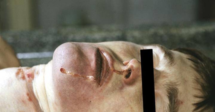
General signs of death from asphyxia. During an external examination of the corpse, a bluish appearance of the face is observed, especially pronounced in the first hours after death; after a few hours it may disappear due to the flow of blood into the underlying sections. Sometimes there is dilation of the pupils, bleeding from the nose, pinching of the tip of the tongue between the teeth and foam at the mouth. Along with this, pinpoint hemorrhages may be found on the inner surface of the eyelids. They can be seen by pulling the eyelids up and down. Minor hemorrhages can also be observed on the skin of the face and neck. Cadaveric spots, as a rule, are very intense, dark purple, and appear quite quickly. Cadaveric spots are a peculiar coloring of the skin of a corpse that forms shortly after cardiac arrest. When blood circulation stops, liquid blood flows down and permeates the underlying tissues and skin of the corpse, giving them the appropriate color. Traces of involuntary urination and defecation are often found in the crotch area of the corpse or on clothing. In men, traces of sperm may be found, released in the form of a drop from the urethra.
During an internal examination of a corpse, there are no diagnostic signs strictly specific for mechanical asphyxia, but the combination of a number of them may be characteristic of death from asphyxia. One of the constant signs is dark, liquid blood. However, it can be detected not only with mechanical asphyxia, but also with rapid death from other causes. With mechanical asphyxia, there is always an overflow of blood in the right half of the heart. The next significant symptom is congestion of the internal organs due to stagnation of blood in the venous system. Often with asphyxia there are small, the size of a millet grain, hemorrhages on the surface of the lungs or between their lobes, under the epicardium, under the mucous membrane of the mouth and upper respiratory tract. These hemorrhages are called ecchymoses, or Tardieu spots. They are formed due to overflow of blood and rupture of the smallest blood vessels. A variable sign is a contracted and anemic spleen.
Hanging
When hanging, death occurs due to compression of the neck by a noose tightened by the weight of the body.
Loops According to the mechanism of their tightening on the neck, they can be stationary or sliding.
The loop has a knot, a ring and a free end. If the knot is tightly tied and the ring does not change its size, then such a loop is called motionless, or fixed. More often, the loop at one end has a small “eye” - a knot; the other free end is threaded into it, as a result of which an easily movable ring of the loop is formed. Such a loop is called a sliding loop.
Depending on the material used for the loops, they are divided into hard (wire, electrical cord, etc.); semi-rigid (belts, thick and coarse ropes) and soft, made of wide soft material, such as a towel, sheet, scarf.
Based on the number of turns of the loops around the neck, loops are divided into single, double, triple and multiple.
In all cases of hanging, when examining the scene of the incident, the noose and its knot must be preserved, since the material of the noose and the method of tying it in some cases can help establish the profession of the victim or murderer (weaver, sailor, fisherman, etc.).
During an external examination of a person who died from hanging or strangulation with a noose, a characteristic and reliable sign is a depressed mark on the neck - strangulation groove.
Rice. 19. Typical (A) and atypical (B) loop application
When a strangulation groove is detected, it is necessary to pay attention to its general appearance, location and direction. Based on these signs, one can judge the nature of the compression of the neck that caused death (hanging or strangulation with a noose). The direction of the groove depends on the way the loop is placed around the neck. When hanging, the noose can be located typically or atypically. A typical strangulation groove is considered when the loop node is located at the back of the head. With an atypical strangulation groove, the loop node is located under the chin or on the side (Fig. 19).
.jpg) Rice. 20. Self-hanging. Hard strangulation groove
Rice. 20. Self-hanging. Hard strangulation groove When hanging, the strangulation furrow is always directed obliquely - from bottom to top. This is caused by the fact that one part of the loop (the free end) is attached to some object (nail, door frame, branch, etc.), and the other, the loop itself, is pulled down by the weight of the body. In this case, the greatest depression of the groove is formed on the side of the loop opposite the node, i.e., in the place of greatest pressure on the neck.
The strangulation groove can be closed, when both ends of it converge in the place where the loop node was located, or open, when the ends do not close with each other.
Depending on the material of the loop, the groove can be hard, soft or transitional. A hard strangulation groove is formed when a loop of dense material with a small cross-section is applied (twine, wire, electrical cord). It is always well defined, depressed, has a parchment appearance, dark brown color, and is dense to the touch (Fig. 20). On corpses and in persons released from the loop and surviving, such strangulation grooves persist for a very long time.
The soft furrows are not clearly expressed and have the appearance of wide, pale bluish, slightly depressed stripes. They are unstable and disappear relatively quickly.
Transitional, or mixed, strangulation grooves are a combination of the first two.
The strangulation groove is a negative imprint of the material of the loop, reflecting its characteristic features: width, the presence of nodes, etc., and is better expressed the longer the corpse was in the loop (Fig. 21).
It is very important for the preliminary investigation authorities to establish whether the groove was formed during the life of the victim or posthumously, since murders with subsequent hanging of the corpse to simulate suicide are known. The intravital strangulation groove is pale and anemic due to the squeezing out of blood from the vessels. Above and below the groove, the vessels are dilated and filled with blood, and small hemorrhages are found in places. The strangulation groove, depending on the number of loops on the neck, can be single, double or multiple. In these cases, raised skin ridges form between its turns. From compression and rupture of small vessels in these ridges, pinpoint hemorrhages occur - this is an almost indisputable sign of intravitality. However, sometimes death in the loop can occur very quickly from cardiac paralysis, and then there may be no blood filling of the vessels and hemorrhages in the area of the groove.
.jpg) Rice. 21. Self-hanging. Strangulation groove from a loop of a belt stitched with patterns
Rice. 21. Self-hanging. Strangulation groove from a loop of a belt stitched with patterns In cases where the corpse of a person strangled by a noose is then hung up to simulate suicide, two strangulation grooves are formed on the neck - one of them is horizontal, intravital, the other oblique, formed posthumously.
To establish whether the furrow is alive, it must be examined in transmitted light. To do this, the skin with the strangulation groove is separated from the soft tissues and examined under the light. If the groove is intravital, then dilated and blood-filled vessels, and sometimes small hemorrhages, are visible along its edges. Along with examination in transmitted light, a binocular stereoscopic microscope can also be used; pieces of the strangulation groove must be examined histologically.
In addition to the strangulation furrow, there are other characteristic signs of death from asphyxia. If the corpse has been hanging in a noose for a long time, then the cadaveric spots are most pronounced on the lower parts of the body and lower extremities. Sometimes pinpoint hemorrhages are visible against the background of the spots. The forearms and hands have a bluish tint. In some cases, compression of the neck by a loop is accompanied by damage to the larynx: fractures or fractures of the large horns of the hyoid bone and the upper horns of the thyroid cartilage are more often observed. Due to the pressure of the loop, hemorrhages occur in the neck muscles. Hemorrhages and even muscle tears at the insertion of the sternoclavicular muscles with the formation of small blood clots may be observed, which undoubtedly indicates that these injuries occurred intravitally. Due to the strong stretching of the neck by the weight of the hanging body, the carotid arteries are simultaneously stretched, which leads to a transverse rupture of their internal membranes below the loop. In this case, an accumulation of coagulated blood can be seen between the stratified membranes. This sign indicates that the damage is alive, but it does not always occur.
It is very important for the investigative authorities to establish whether there are any injuries on the corpse.
In such cases, the forensic medical expert must determine the nature of these injuries - intravital or postmortem - and how they were caused.
.jpg) Rice. 22. Hanging on the collar of clothes. Alcohol intoxication. Accident (personal observation)
Rice. 22. Hanging on the collar of clothes. Alcohol intoxication. Accident (personal observation) In most cases, hanging is a suicide, but accidents and even murders can occur. In suicides, the body of a hanged person during convulsions may hit nearby hard objects, such as protruding parts of rooms, door frames, metal parts of stairs, wood knots, etc. In these cases, the damage is superficial and is located on protruding parts of the body - on the nose , chin, hands. Self-hanging may also reveal more severe injuries, including cut and stab wounds that were inflicted for the purpose of suicide before hanging. This is often observed in mental patients.
When killed by hanging, the damage to the corpse is intravital in nature. In such cases, as a result of struggle and self-defense, hemorrhages and abrasions occur on the arms, neck, face, and chest. Cases of murder without any damage are possible when, by deception or during sleep, a noose is placed around the neck, and its free end is quickly secured to some object.
It is almost impossible to decide whether there was a murder, accident or suicide based solely on the nature of the injuries. It is necessary to carefully examine the scene of the incident, its setting, the posture and clothing of the hanged person, the nature of the noose and knots, as well as all changes on the corpse.
Circumstances of the hanging. In most cases, self-hanging is committed by people who are mentally unstable or in a state of alcoholic depression. There may be cases of suicide among school-age children due to various childhood experiences and other motives.
As already noted, with hanging there may be cases of murder by fraudulently placing a noose around the neck of a physically healthy person or someone in a state of sleep. Murders are possible by hanging the sick, the physically weakened, and those in a state of heavy alcoholic intoxication. The presence of a large amount of alcohol in the internal organs during death from hanging may indicate murder, since in a state of severe alcoholic intoxication such persons not only cannot resist, but are also unable to commit self-hanging on their own.
.jpg) Rice. 23. Self-hanging: legs bent, touching the ground
Rice. 23. Self-hanging: legs bent, touching the ground Accidental self-hanging may result in an imitation or simulation of suicide.
Hanging as an accident is rare. Its victims are mainly small children: a child sticks his head between the bars or into a torn bed net, in which his neck is pinched, loses consciousness and dies from asphyxia. A similar death due to accidental pressure of the neck against any narrow object occurs in adults who are intoxicated. In our practice, there was a case when a drunk sat down on the steps of the porch, and on the railing hung rope reins rolled up in several turns, into which he stuck his head and fell asleep. Due to the pressure of the neck on the reins, self-hanging and death occurred,
In another case, a man in a state of intoxication, wanting to warm up, sat on a stool near the stove door and, while falling asleep, his jacket collar caught the protruding end of the door latch. Death occurred from compression of the neck by a clothing collar (Fig. 22),
.jpg) Rice. 24. Paired self-hanging
Rice. 24. Paired self-hanging Poses of the hanged, in particular, suicides are very diverse: hanged people can hang freely in a noose; the head can be in a loop, and the legs in a half-bent or bent position rest on the floor, on the ground (Fig. 23). Suicides can be in various positions: standing on their feet, kneeling, in a reclining or lying position on the bed, while the free end of the loop can be secured to the headboard or another object. Sometimes a suicide person, before hanging himself, puts on a noose and ties his legs or arms, or both. There are also paired suicides, when a man and a woman commit suicide in the same loop (Fig. 24).
Asphyxia caused by exposure to a mechanical factor on the body is called mechanical asphyxia. The concept of “asphyxia” is translated as “lack of pulse” (a - negation, sphygmos - pulse). Mechanical asphyxia is based on mechanical obstacles to the entry of air into the lungs. In the genesis of such asphyxia, two main factors play a role: acute oxygen deficiency and the simultaneous accumulation of carbon dioxide, which determines the occurrence of the pathophysiological process.
With mechanical asphyxia, the access of air into the body through the respiratory tract is stopped, and therefore oxygen is quickly consumed by the tissues and carbonic acid accumulates in them. Within a few minutes, this leads to paralysis of the central nervous system and death. Thus, mechanical asphyxia is mainly characterized by: the action of an external factor that mechanically interrupts the circulation of air in the respiratory tract, and as a consequence of this, the almost complete disappearance of oxygen from the blood and tissues and the accumulation of carbon dioxide in them.
Classification:
1. strangulation asphyxia:
- hanging;
- strangulation with a loop;
- manual strangulation;
- strangulation with a hard object
2. obstructive asphyxia:
- closing the openings of the mouth and nose with hands and soft objects;
- closing the airway lumen with compact foreign bodies;
- aspiration of bulk solids
- aspiration of liquids
- aspiration of gastric contents
- drowning in water:
- true ("wet")
- asphyxial (“dry”)
- drowning in other liquids
- compression asphyxia: compression of the chest and abdomen;
- asphyxia in a limited confined space.
Distinguish 7 stages of asphyxia: 1) pre-asphyxial, 2) inspiratory dyspnea, 3) expiratory dyspnea, 4) short-term respiratory arrest (or rest period), 5) terminal breathing, 6) persistent respiratory arrest. 7) cardiac arrest.
The first pre-asphyxial stage. This stage usually lasts the first 10-20 seconds, but can last several minutes. A person’s training in holding their breath plays a big role here.
The period of inspiratory dyspnea. During this stage, usually lasting about 1 minute, inhalation prevails over exhalation. This phase depends mainly on the volume of the lungs and the amount of air in them. Depletion of blood in oxygen and accumulation of carbonic acid reflexively and directly irritate the central nervous system and cause the onset of shortness of breath that increases in depth and lengthens the respiratory rhythm.
The third stage is the period of expiratory dyspnea, in which exhalation prevails over inhalation. This stage is manifested by contraction of the body muscles up to muscle cramps. At this stage, blue mucous membranes also occur, dilation of the pupils, slowing of the heartbeat, first with an increase and then a decrease in blood pressure. In the second minute, breathing at the height of inspiration is interrupted by single convulsive twitching of individual muscle groups, presumably from irritation of the corresponding areas of the cortex. At the end of the first - beginning of the second minute, consciousness is lost; By about the third minute, irritation spreads to the entire cortex, and general convulsions occur with the release of feces and urine. Convulsions end in opisthotonos.
The fourth stage of asphyxia is rest. This stage lasts for several seconds or minutes.
After 30 - 45 seconds from the beginning of the resting stage, individual rare and weak contractions of the respiratory muscles appear - “terminal breathing” - the fifth stage; heart contractions become more frequent but become weaker. By the end of the fourth minute, terminal breathing freezes, only a gradually weakening heartbeat remains.
The sixth stage of asphyxia is the final cessation of breathing.
The seventh stage is cardiac arrest, which occurs in the 5th to 8th minute.
The intensity of the severity and duration of individual stages of asphyxia depend to a certain extent on a number of factors: the type of mechanical asphyxia, age, state of health, etc.
Mechanical asphyxia is accompanied by severe disorders of the central nervous system. Consciousness is lost at the end of the first or at the beginning of the second minute; during strangulation, especially when hanging, much earlier. With slowly developing asphyxia, loss of consciousness is preceded by visual and hearing disturbances, and the sense of pain is lost.
Mechanical asphyxia is characterized by rapidly onset adynamia, active movements become impossible. Increased excitability of the smooth muscles of the intestines and bladder while relaxing the sphincters leads to involuntary eruption of urine and feces. For the same reason, seminal fluid is released in men and the contents of the cervical canal in women.
Signs of asphyxia:
External signs:
small hemorrhages in the connective membrane of the eyes - can be multiple, most often localized in the transitional folds of the conjunctiva; with long-term asphyxia, the same hemorrhages can form in the skin of the eyelids, face, neck, upper chest, and on the mucous membrane of the mouth; this sign, indicating an increase in intravenous pressure and an increase in the permeability of the vascular wall due to hypoxia, is valuable, but it is not constant.
facial cyanosis is a common but also unstable sign that can disappear in the first hours after death as a result of blood flowing into the underlying parts of the corpse; on the other hand, when the corpse is positioned face down, cyanosis can also occur in cases where death is not associated with mechanical asphyxia.
diffuse intense dark purple cadaveric spots - their intensity is associated with the liquid state of the blood and therefore its easy movement to the underlying parts of the body; this state of cadaveric spots is typical for all cases when death occurs quickly, therefore it is typical for all cases when death
occurs quickly, so the diagnostic value of this symptom is small;
involuntary urination, defecation and eruption of sexual secretions - are not observed in mechanical asphyxia in every case and are sometimes observed in other types of death (electrical trauma, poisoning with certain poisons, sudden death).
Small hemorrhages in the connective membranes of the eyes, less often in the skin of the face, neck and oral mucosa are a valuable sign of asphyxia. These hemorrhages can be numerous or isolated, most often localized in the transitional folds of the conjunctiva. They are formed as a result of increased pressure in the superior vena cava system and increased permeability of the vascular wall due to hypoxia. Congestion and cyanosis of the face occur already in the first minutes of the asphyxial process and often persist on the corpse, but often disappear several hours after death as a result of partial drainage of blood into the lower parts of the corpse. Slower cooling of the corpse, other specified equal conditions, rapid formation of diffuse, intense cadaveric spots, rapid rigor mortis, rapid onset of putrefaction, discharge of urine, feces, sperm.
Internal signs:
- dark liquid blood is a sign constantly observed during mechanical asphyxia; however, the same state of the blood is characteristic of many other types of quickly occurring death; The dark color of the blood is explained by the post-mortem absorption of blood oxygen by surviving tissues.
- overflow of blood to the right half of the heart - associated with difficulty in blood circulation in the pulmonary circle; in case of rapid death, there is always more blood in the right half of the heart than in the left; however, in death from mechanical asphyxia, the difference in the blood supply of both halves of the heart is always more distinct.
- congestion of internal organs - occurs in many types of rapid death; in itself it has no diagnostic value.
- relative anemia of the spleen is a relatively rare symptom; It is assessed differently by different authors, but the majority is inclined to believe that anemia of the spleen in combination with other data should be used to diagnose death from mechanical asphyxia.
- subpleural and subepicardial small hemorrhages are a common finding in mechanical asphyxia. Their size is usually small - from pinpoint to the size of a millet grain, the color is intensely dark red, often with a bluish tint; their number ranges from single to ten or more; under the pleura of the lungs they are most often found on the diaphragmatic and interlobar surfaces, on the heart - under the epicardium on its posterior surface; the occurrence of these hemorrhages is due to a sharp increase in pressure in small veins and the capillary network during the period of convulsions, as well as an increase in the permeability of the vascular wall as a result of oxygen starvation of tissues; minor hemorrhages during mechanical asphyxia are observed not only under the serous membranes, but also in the muscles and in all internal organs, as a morphological manifestation of an extremely rapid reaction of the vascular system to the occurrence of acute oxygen starvation in the body; small hemorrhages under the pleura and epicardium also occur in other types of death, but with mechanical asphyxia they are more common and more numerous.
- acute alveolar, less often interstitial, pulmonary emphysema.
A liquid state of blood in the heart and blood vessels of a corpse, caused by hypercapnia, is constantly observed in cases of death from mechanical asphyxia. The liquid state of the blood leads to the rapid formation of intense confluent cadaveric spots. Overflow of blood to the right side of the heart is associated with stagnation and hypertension in the pulmonary circulation. Small hemorrhages /ecchymoses/ in the pleura and epicardium (Tardier spots) are a common finding in mechanical asphyxia - their edges are clear, intense, dark red, sizes range from dotted to 1-2 mm. in diameter, number from single to multiple, most often found on the posterior diaphragmatic surface of the lungs, in between the lobar fissures, on the posterior surface of the heart. The occurrence of these hemorrhages is caused by a sharp increase in pressure in the capillaries and venules, an increase in the permeability of the vascular wall due to hypoxia, as well as a drop in pressure in the pleural cavities during the stage of inspiratory dyspnea. Such hemorrhages sometimes occur not only in the serous membranes, but also in the muscles, internal organs, and mucous membranes of the gastrointestinal tract. They are a morphological sign of an extremely rapid response of the vascular system to the occurrence of oxygen starvation. In the lungs there is emphysema of varying degrees (most pronounced in drowning).